

Biopharmaceutical products span a vast size range, from proteins and antibodies at the nanometre scale through to microparticles in cell-based therapies. Regulatory attention focuses most acutely on the subvisible microparticle range, roughly 1 to 100 micrometres, because particles in this range can occlude small blood vessels in sensitive organs including the brain and lungs. The relationship between particle characterisation, quantification and patient risk is direct, and regulatory agencies expect sponsors to demonstrate they understand it. The pharmacopoeial framework for subvisible particle analysis specifies three principal methods in USP chapters 787, 788 and 789: membrane microscopy, light obscuration and flow imaging. Each has a defined scope, established acceptance criteria and specific limitations. A sponsor who selects the wrong method for their product type, or who validates it inadequately, risks both failing specification limits and generating data that cannot be defended in a regulatory submission. The decision is not simply technical. It is strategic.
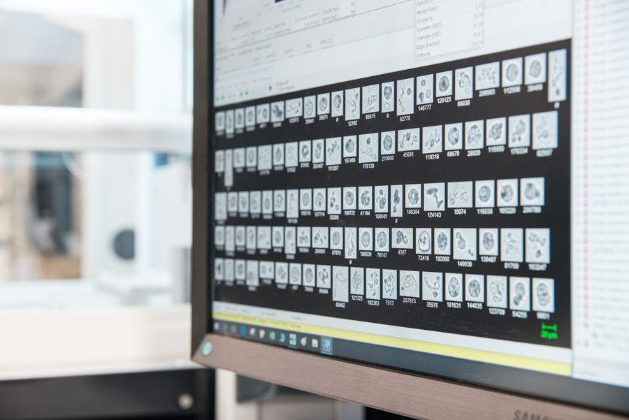
Microflow imaging particle classification display, Coriolis Pharma analytical laboratory.
Light obscuration has been the established method for subvisible particle analysis for over 40 years. It operates by detecting the drop in light intensity as particles pass through a laser beam, generating a count and size distribution. Its longevity means that its analytical procedures are well described in pharmacopoeias and its regulatory acceptance is broadly understood. For many conventional biologics, particularly those with relatively opaque particles and sufficient sample volumes, it remains an appropriate first-line method.
Microflow imaging takes a fundamentally different approach. Rather than measuring light obstruction, it captures high-resolution images of individual particles as they pass through a flow cell, classifying each by morphological properties including circularity, aspect ratio and edge gradient. The practical advantages over light obscuration are significant. Sample volume requirements are lower, typically 0.5 millilitres against 2 to 5 millilitres for light obscuration. Sensitivity for transparent and semi-transparent particles is substantially higher. And crucially, the morphological data allows particles to be discriminated by type: proteinaceous aggregates, silicone oil droplets and foreign particulates can be distinguished in a way that light obscuration simply cannot achieve. For highly concentrated formulations, for low-volume clinical candidates and for products where particle identity matters as much as particle count, microflow imaging is the more informative method.The choice between them is not binary. A well-designed analytical strategy may use both in combination, applying light obscuration for routine batch release where speed and regulatory precedent matter, and microflow imaging for formulation development and root cause investigations where morphological classification adds the most value. What matters is that the selection is deliberate, documented and grounded in the specific attributes of the product being developed.
“Microflow imaging offers significantly lower sample volume requirements and higher sensitivity for transparent particles. It also provides crucial morphological information that helps discriminate particle types and identify their sources.”
Dr Bodo Brocks, Head of Quality, Coriolis Pharma
The transition from research-grade analytical methods to validated GMP procedures is one of the most practically demanding steps in biopharmaceutical development, and subvisible particle analysis is a particularly common source of difficulty. Methods developed under R&D conditions are often optimised for sensitivity and information density rather than robustness, reproducibility and regulatory acceptability. When these methods are transferred to a GMP environment without adequate bridging work, the result is data that cannot support a regulatory submission and a validation programme that must be substantially redesigned under time pressure. Sample preparation is the most frequent source of failure. Subvisible particle counts are highly sensitive to how samples are handled before measurement. Agitation, temperature excursions, contact with incompatible surfaces and inadequate degassing can each introduce artefactual particles that inflate counts or mask genuine particle populations. Establishing standardised sample preparation procedures, validating them as part of the analytical method and documenting the controls that prevent contamination are prerequisites for GMP-quality data. These controls cannot be retrofitted after the fact without repeating a substantial portion of the validation work.Method validation for subvisible particle analysis must address specificity, precision, accuracy, range and intermediate precision as a minimum. For microflow imaging specifically, the classification thresholds used to discriminate particle populations must be defined and justified. Acceptable limits must be set before validation commences, not adjusted post hoc to accommodate results. Regulatory reviewers are experienced at identifying validation packages where acceptance criteria appear to have been set to fit the data, and such packages invite additional scrutiny or requests for repeat studies. Coriolis Pharma’s analytical development team supports sponsors through this process, having established validated GMP methods for subvisible particle analysis across a range of biologic modalities, from conventional monoclonal antibodies to complex biologics and gene therapy vectors.Coriolis Pharma is a Munich-based CRDO specialising in biologics drug product development, with particular expertise in formulation development, analytical characterisation and lyophilisation. The organisation supports programmes across a broad range of biologic modalities and development stages, from early feasibility through GMP clinical manufacture, and maintains analytical capabilities that span the full spectrum of subvisible particle characterisation methods described in current pharmacopoeial guidance.
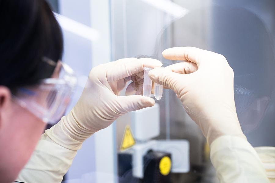
Sample handling in controlled GMP environment, Coriolis Pharma.
“We live in a world of particles. Regulatory authorities focus on microparticles because they can cause blockages in small blood vessels. The interplay between characterisation and quantification is what determines patient risk.”
Dr Bodo Brocks, Head of Quality, Coriolis Pharma
Watch the webinar on demand The full presentation on subvisible particle analysis, covering method selection, validation strategy and the R&D to GMP transition in detail, is available on demand via the Pharma D-mand webinar library. For organisations building out their particle characterisation programmes, the Pharma D-mand advisory team can connect you with the relevant expertise.